Human Stem Cell Culture (High Concentration Human Stem Cell Culture Dry Freeze) What is Dry Freeze?
1. AT high-concentration human stem cells (100HMSC-AT) adipose tissue mesenchymal stem cells The mesenchymal stem cells that exist in bone marrow and adipose tissue are cells that can become "seeds" for adding new cells. Stem cells for body repair and regeneration decrease with age. When the number of stem cells is reduced, the repair of the tissue will be too late for aging. By replenishing this reduced stem cell, the injured part and the aging part are repaired, and the whole body is rejuvenated. Adipose stem cells have two characteristics: "self-replication ability" and "differentiation ability". As adult stem cells with the same characteristics, there are umbilical cord blood, placenta, bone marrow, especially adipose stem cells. They are easy to collect and can collect a large number of stem cells, so they are attracting attention. Once the adipose-derived mesenchymal stem cells are removed from the body, the secretion of growth factors becomes active. After returning to the body, it differentiates into bone cells, chondrocytes, fibroblasts, skeletal muscle cells, fat cells, etc., and the secreted growth factors also have a good effect on the growth of other cells.
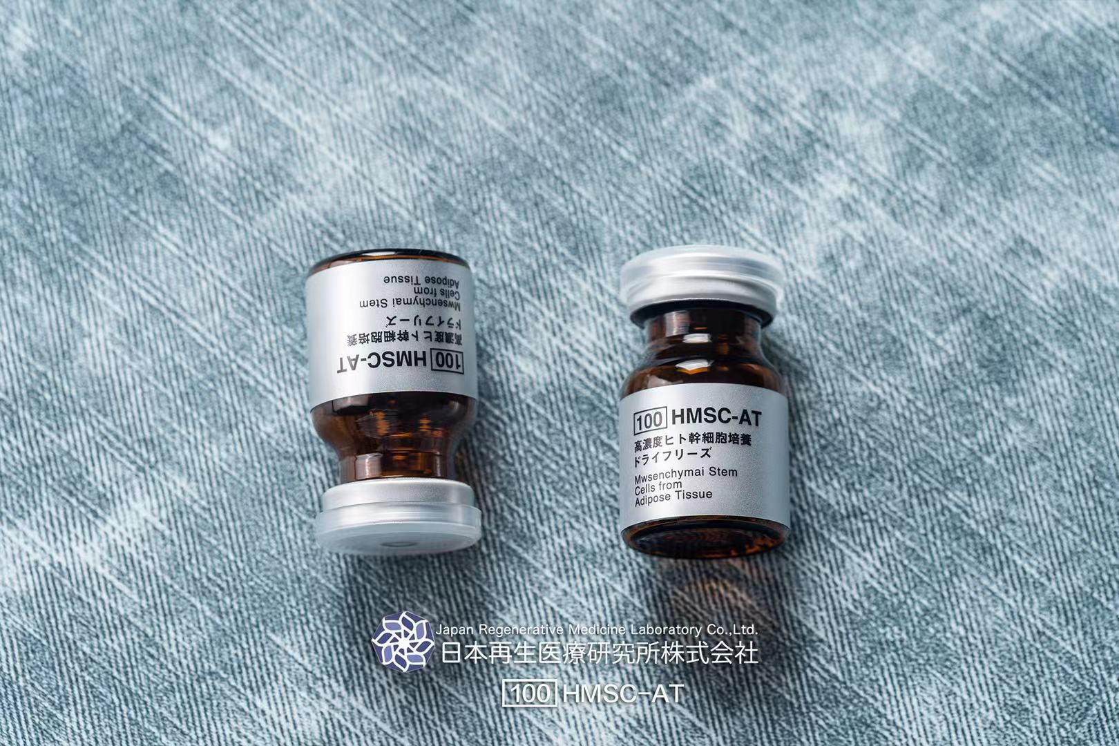
2. AT high concentration human stem cells (100HMSC-AT) High-concentration
pit stem cell culture, anti-aging and anti-aging are not only women’s business!
Men's anti-aging should not be missed! Stem cell anti-aging can rejuvenate
the body's functions from the inside to the outside through the intrinsic
strength of the body, so that the renewal of senescent cells tends to be
normal, so as to achieve the purpose of anti-aging. Using stem cells (100HMSC-AT)
for the first time clearly felt the comprehensive improvement of the functions
of the body's various systems, which can be used for beauty, anti-aging,
sleep aid, and prevent potential diseases. Improve the renewal speed of
body cells, the human body is full of energy and vitality, and naturally
becomes younger and younger, which fundamentally achieves the purpose of
delaying aging and longevity! AT high concentration of human stem cells,
a gas station for youth
3. AT high-concentration human stem cells (100HMSC-AT) are derived from human adipose tissue mesenchymal stem cells, and cultivate the dental pulp, umbilical cord, bone marrow, fat and other stem cells classified as mesenchymal stem cells in the human body. When the number of cells is increased, the second-generation solution is called "human-derived stem cell culture supernatant". After removing the stem cells from the stem cell culture solution, the liquid (supernatant) that was subjected to various sterilization treatments. This human stem cell culture supernatant contains the same cell delivery agent as the stem cells. The same effect as the stem cells was observed, and clinical trials started. Medical use. The effect is also confirmed. AT high concentration of human stem cell effects: 1. Anti-aging, skin becomes smoother, moisturized, skin tone becomes whiter, fine wrinkles become lighter, facial spots become lighter, skin becomes firmer all over, breasts and buttocks become firmer and more elastic 2. Improve immunity, improve sleep, less fatigue, energetic3, promote blood circulation, anti-oxidation, alleviate women's menopausal symptoms4, comprehensively improve human body functions, regulate hormone levels in the body, increase metabolism Speed up the healing of wounds!
4. 100HMSC-AT (High Concentration Stem Cell Culture Dry freeze) is derived
from human adipose tissue mesenchymal stem cells, which can use stem cells
to differentiate into other cells in the body, regeneration and re-fixation
to repair various degenerative, necrotic, invasive, Metabolism and degenerative
disease, restore diseased or degenerated tissue and organ structure, and
have this unique function. Repair and replace various aging and damaged
tissue cells, repair the origin of muscle basic cells, restore the youthful
physiological ability of the cells, make the cells have lasting youthful
vitality, and fade stains from the inside out, smooth fine lines, and skin
Restore softness, restore firmness, whiten and brighten skin. Stem cell
beauty and anti-aging. From a medical point of view, anti-aging is a holistic,
integrated and in-depth medical behavior, not just the face, but only a
full range, three-dimensional, deep anti-aging, and achieving the best
anti-aging effect from the inside to the outside. The body is full of vitality,
improves immunity, and improves facial problems.
5. AT high concentration of human stem cells (100HMSC-AT) There are collagen, hyaluronic acid, elastin, etc. in skin regenerative medicine. They are all basic elements such as elasticity, moisturization, and elasticity. These are all made by skin cells and work to keep young. In normal life, skin cells are damaged by stress and ultraviolet rays. And with age, the function of the skin will decline and the number will decrease. If skin cells decline, the essential elements of the skin will not be produced, which can cause wrinkles and sagging. Transplanting "fibroblasts", "PRP", etc. onto the damaged skin can play a role in repairing the weakened skin tissue. As a result, collagen, hyaluronic acid, elastin, etc. are reproduced, and the skin tissue is improved. The skin itself can restore its original youth. Unlike the treatment of injecting foreign bodies such as hyaluronic acid, it will not be absorbed by the body and can delay the aging rate for a long time. 6. I sincerely recommend the 100HMSC-AT high-concentration human stem cell culture medium for personal use: stem cells are known as the "universal cells" in the medical field. It can inhibit inflammation and allergies, improve rough skin, dilute acne marks, whiten spots, improve wrinkles and skin elasticity, regulate endocrine and repair aging cells. Fundamentally achieve the effects of anti-aging and anti-aging! Applicable people: 1. Prevent aging, dull skin, sagging, facial beauty 2. Endocrine and sexual dysfunction 3. People with symptoms such as low immunity, body aches 4. Insufficient sleep, poor mood, deterioration of body function 5. Slow metabolism , Menstrual disorders, etc.
6. Prevent disease, anti-virus, etc. It is recommended to activate cell
regeneration, promote collagen production, and increase skin gloss and
elasticity once a month (the effect is best)! AT high-concentration human
stem cells are derived from high-end medical anti-aging products in Ginza,
Japan. They secrete cellular nutritional factors to achieve nutrition and
repair of healthy cells, thereby achieving a comprehensive repair of the
human body. AT high concentration of human stem cells makes every cell
of the body, every inch of skin, every organ, and every second of spirit
full of youthful vitality. To keep us lasting: health, youth and beauty!
What is stem cell therapy (derived from autologous fat)?
What are stem cells?
We all have the ability to regenerate and replenish lost cells in our bodies to keep individual cells, like skin and blood, short-lived and constantly changing tissues. I have cells. Cells with these abilities are "stem cells." To be called a stem cell, the following two abilities are indispensable. One is the ability to produce various cells that make up our body, such as skin, red blood cells, and platelets (differentiation ability), and the other is the ability to divide into cells with exactly the same ability as ourselves (ability to divide). Self-replicating ability).
Stem cells can be broadly divided into two types. One is stem cells, such as skin and blood, that continue to make replacement for disappeared cells in fixed tissues and organs. This type of stem cell is called a "tissue stem cell". Tissue stem cells can not be anything, but the role is decided such that hematopoietic stem cells that make blood are blood system cells, and neural stem cells that make nervous system are only neural cells. The other is a "pluripotent Stem Cell" that can be produced by any cell in our body, such as ES cells (embryonic stem cells). In other words, pluripotent stem cells can also produce various tissue stem cells in our body. iPS cells (induced Pluripotent Stem Cells) are "pluripotent stem cells" that are artificially created from ordinary cells.
Utilizing these properties of "stem cells", research on a new treatment method called "regenerative medicine" that cures injuries and illnesses using the cells themselves as drugs, and the mechanism of illness by reproducing the state of cells in the body outside the body Research is underway to investigate.
MSC (Mesenchymal Stem Cell) Principle
Effect of MSC transplantation on diabetic cardiomyopathy. (A) MSCs increase MMP-2 activation, suppress MMP-9 activation, and attenuate cardiac remodeling. (B) MSCs produce VEGF, IGF-1, AM, HGF and stimulate myogenesis and angiogenesis in injured myocardium. (C) Through differentiation into cardiomyocytes and vascular endothelial cells, MSCs improve myocardial perfusion and myocardial regeneration. Abbreviations: AM, adrenomedullin; HGF, hepatocyte growth factor; IGF-1, insulin-like growth factor-1; MMP, matrix metalloprotease; MSC, mesenchymal stem cells; VEGF, vascular endothelial growth factor
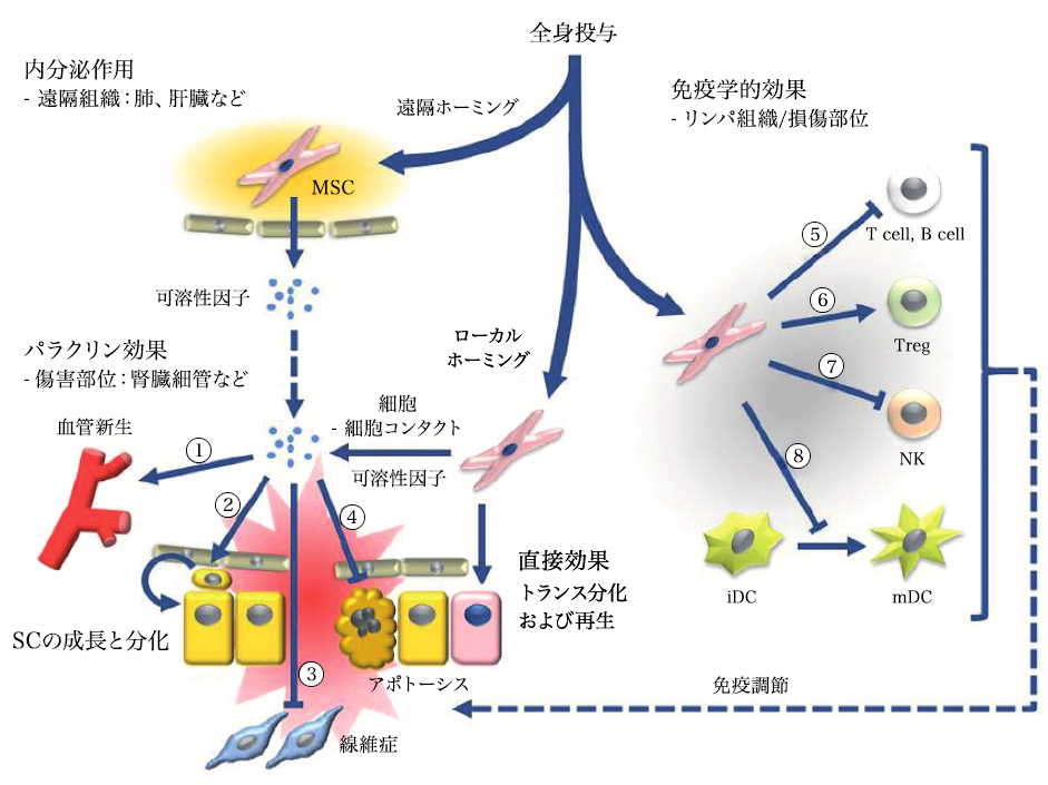
Systemic administration of mesenchymal stem cells can induce terminal (endocrine) or local (paracrine) effects, including cell-mediated effects. 1) Vascular endothelial growth factor (VEGF), insulin-like growth factor 1 (IGF-1), monocytic chemoattractant protein 1 (MCP1), basic fibroblast growth factor (bFGF), interleukin 6 (IL6) 2 ) Stem cell growth and differentiation: Stem cell factor (SCF), Leukemia inhibitor (LIF), Macrophage colony stimulator (M CSF), Stroma cell-derived factor 1 (SDF1), Angiopoietin 1, Activin A 3) Inhibition of fibrosis : Hepatic cell growth factor (HGF), bFGF, adrenomedulin (ADM) 4) Inhibition of apoptosis: VEGF, HGF, IGF1, transformed growth factor (TGF) β, bFGF, granulocyte macrophage colony stimulator (GM CSF), activin A, Thrombospondin 1. Immune-mediated effects include (5-8) 5) T and B cell suppression: human leukocyte antigen G5 (HLA G5), HGF, inducible nitric oxide synthase (iNOS), indoleamine 2 , 3 Dioxygenase (IDO), Prostaglandin E2 (PGE 2), bFGF, TGFβ 6) Induction of differentiation and proliferation of regulatory T cells (Treg) by TGFβ expression. 7) Inhibition of natural killer (NK) cells by the secretion of IDO, PGE 2 and TGFβ. 8) Inhibition of dendritic cell (DC) maturation by PGE 2 secretion.
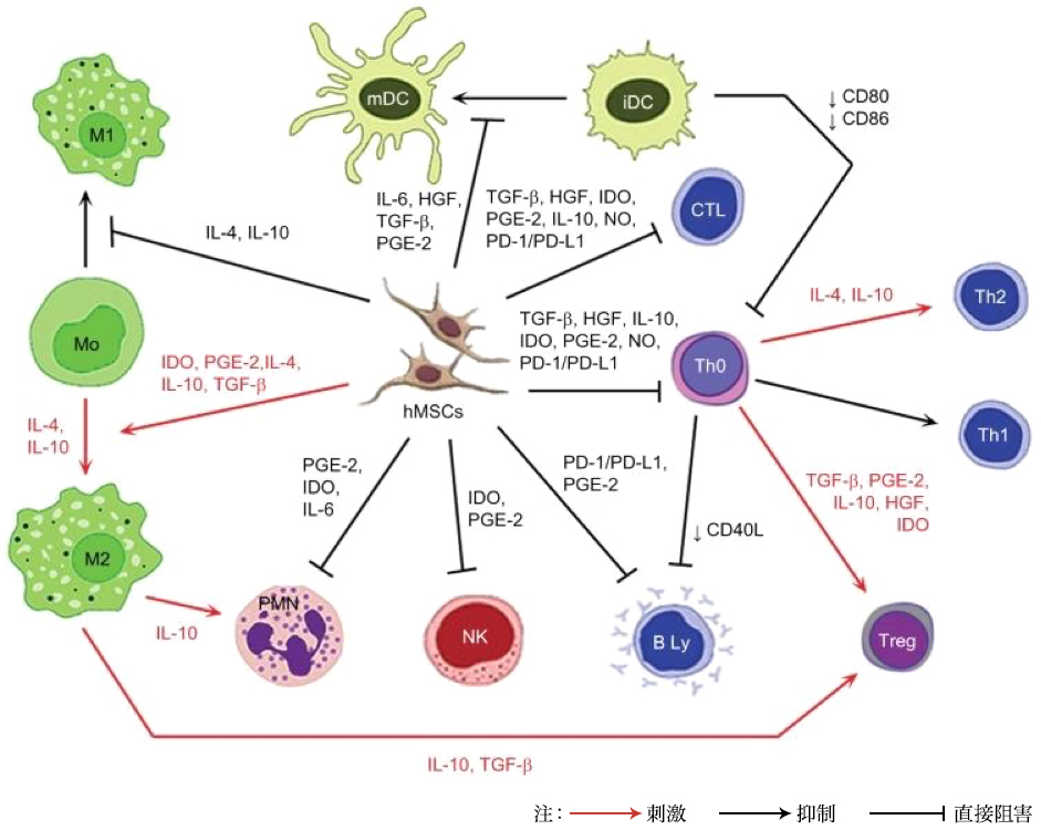
Note: Red arrow: Stimulation; Black arrow: Suppression; Hookless arrow: Direct inhibition
Abbreviations: iDC, immature dendritic cells; IL, interleukin; HGF, hepatocellular growth factor; TGF-β, transformed growth factor-β; PGE-2, prostaglandin E2; IDO, indolamine 2,3- Dioxygenase; NO, nitrogen monoxide; PD-L1, programmed death ligand 1; hMSC, human mesenchymal stem cells; Treg, T regulatory; Th, T helper; CTL, cytotoxic T cells; mDC, mature trees Dendritic cells; PD-1, programmed cell death protein 1; PMN, polymorphonuclear leukocytes; NK, NK cells
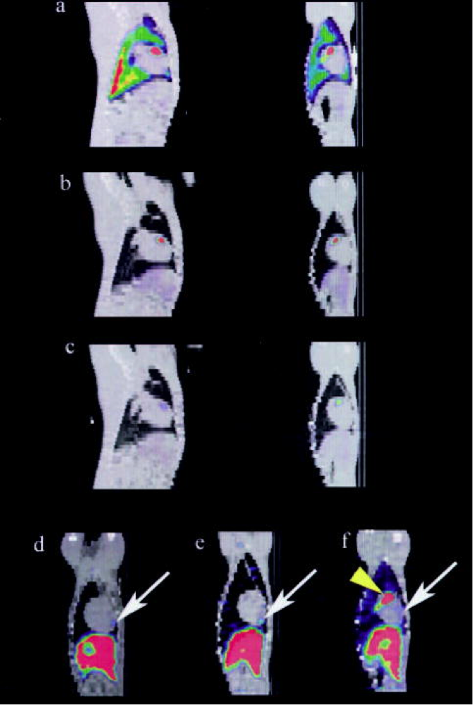
Sagittal plane (left) and SPECT / CT images fused on days 1 (a), 2 (b) and 7 (c) showing local uptake in the anterior ventricular region of the animal heart It is the figure of the coronal plane (right). At the time of the last imaging (day 5-8), the anterior apical region (arrow) of MSC ingestion is shown in the coronary structure diagram for three representative animals. The vertex distribution ahead of this was present regardless of whether early focal hotspots were observed. (Only the yellow arrow of f)
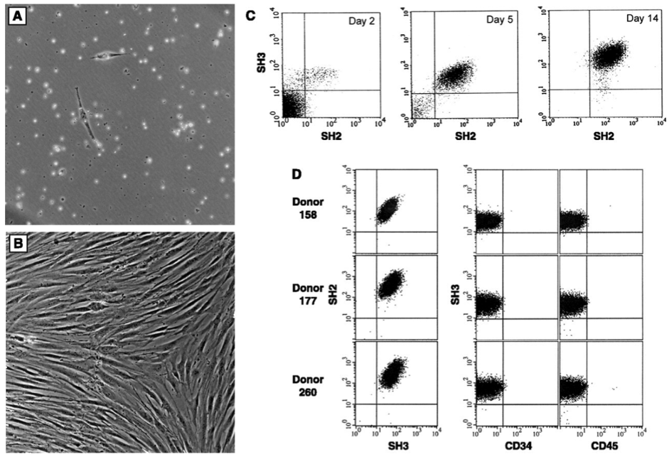
Characteristics of isolated bone marrow stromal cells. Cells are cultured from bone marrow after density fractionation and are shown as (A) 48 hours after plating and (B) 10 days after plating. (C) Flow cell metrology shows enrichment of these cultured cells. Results were obtained on days 2, 5, and 14 of culture using the antibodies SH2 and SH3 generated against surface markers. (11) On day 14, cells were 95-99% homogeneous and negative for the antigens CD14, CD34 (Becton-Dickinson), or CD45 (Pharmingen) common to hematopoietic cells. (D) The homogeneity and reproducibility of the isolation procedure was demonstrated by fluid cell metrology.
MSC (Mesenchymal Stem Cell) Principle
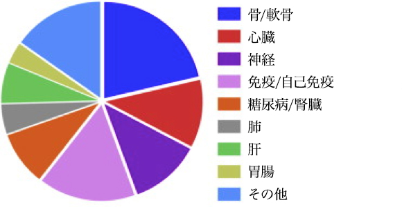
Indications indicated using MSC in clinical trials. Data from 352 registered clinical trials
Indications Being Addressed using MSCs in Clinical Trials. Data for 352 registered clinical trials.
Citation// Stem Cell Therapies in Clinical Trials: Progress and Challenges. Trounson, Alan et al. Cell Stem Cell , Volume 17 , Issue 1 , 11 – 22
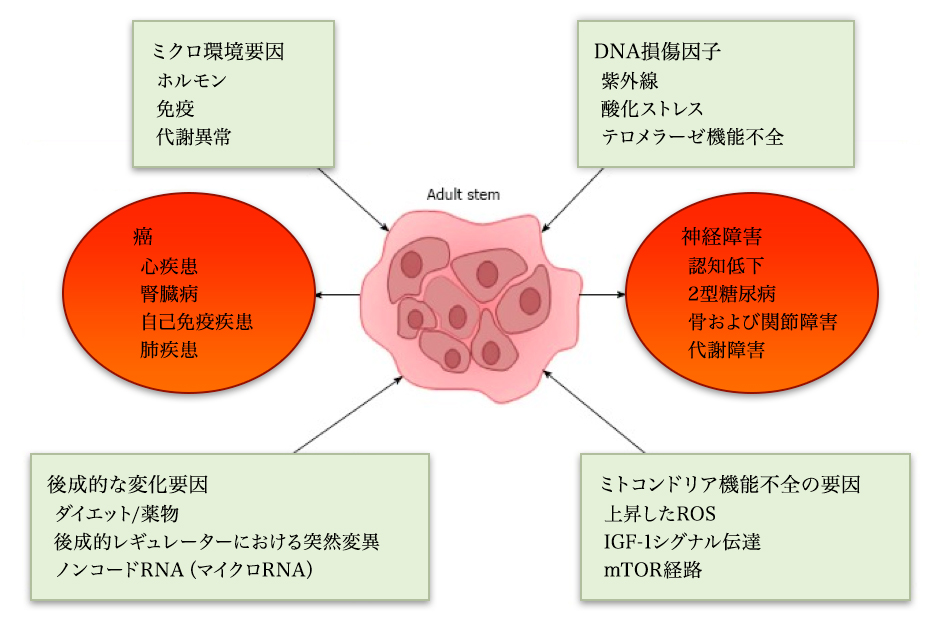
Diseases by disease and age

Pluripotent stem cells have remarkable self-renewal ability and can differentiate into a plurality of diverse cells. There is increasing evidence that the aging process can adversely affect stem cells. As stem cells age, their ability to regenerate declines and their ability to differentiate into various cell types changes. Therefore, it is suggested that the decrease in stem cell function due to aging may play an important role in the pathophysiology of various aging-related diseases. It is important to understand the role of the aging process in stem cell function not only in understanding the pathophysiology of aging-related diseases, but also in developing effective stem cell-based therapies for the treatment of future aging-related diseases. .. This review article focuses on the basis of various aging-related diseases, stem cell dysfunction. Next, we describe some concepts regarding the mechanisms that can cause aging-related stem cell dysfunction. We also briefly discuss current potential therapies for aging-related stem cell defects under development.
By disease / liver / diabetes
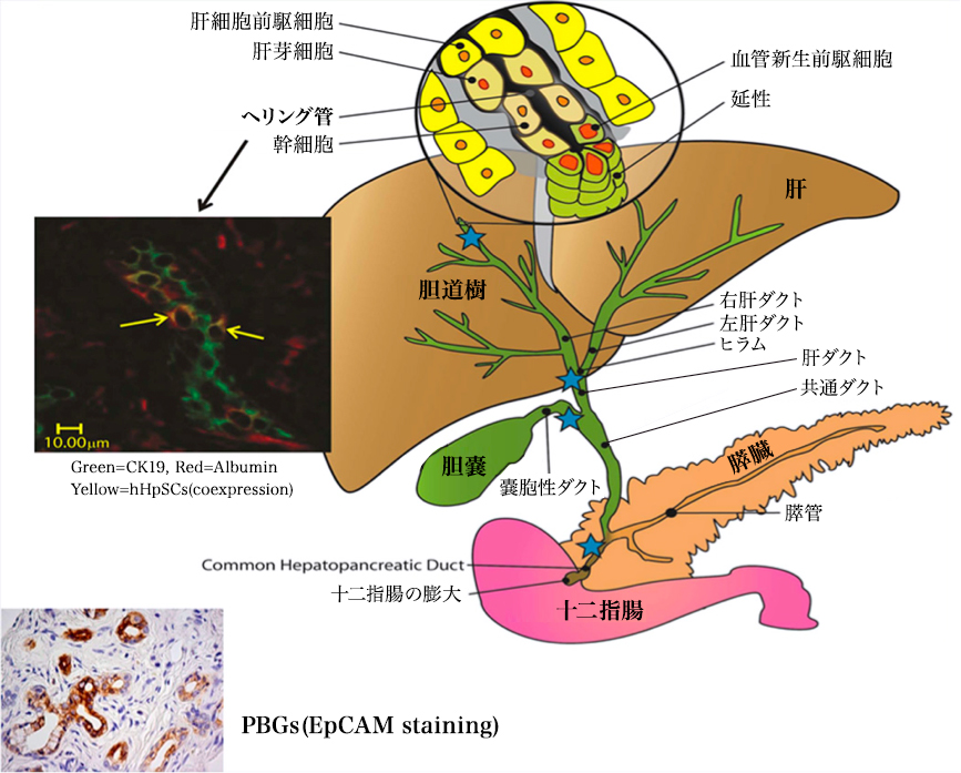
Regenerative medicine is moving to clinical programs with stem cell / progenitor
cell therapies to repair damaged organs.The liver and pancreas, the organ
that shares the endoderm stem cell population, and the biliary tract stem
cells (hBTSC) located in the biliary tract stem are briefly described.
They are the precursors of hepatic stem / progenitor cells in the Herring's
duct and the precursors of the pancreatic duct gland. They give rise to
a mature lineage along the radial axis within the bile duct wall, and a
proximal-distal axis starting from the duodenum and ending with mature
cells in the liver or pancreas. Clinical trials have been conducted for
many years to evaluate the effects of stem cells (fetal liver-derived hepatic
stem / progenitor cells) transplanted into the hepatic arteries of patients
with various liver diseases. Immunosuppression was not required. All control
subjects given the given criteria died within 1 year or had reduced liver
function. Subjects transplanted with 100-150 million liver stem / progenitor
cells showed improved liver function and survival over several years. The
assessment of transplant safety and efficacy is still underdeveloped. Stem
cell therapy for diabetes with hBTSC is still being studied, but is likely
to occur after ongoing preclinical studies. In addition, mesenchymal stem
cells (MSCs) and hematopoietic stem cells (HSCs) have been used in patients
with chronic liver disease or diabetes. MSCs have been shown to be effective
through paracrine signaling of trophic and immunomodulatory factors, limiting
inefficient phylogenetic restriction to mature parenchymal or islet cells.
The effect of HSC is mainly due to the regulation of the immune system.
Stem Cells. 2013 Oct;31(10):2047-60. doi: 10.1002/stem.1457. Concise review: clinical programs of stem cell therapies for liver and pancreas.Lanzoni G1, Oikawa T
Diabetes
QinanWu, Bing Chen, and Ziwen Liang,
Mesenchymal Stem Cells as a Prospective Therapy for the Diabetic Foot
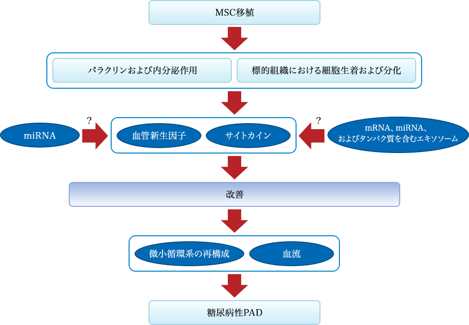
Figure 1: Mechanism of the effect of MSC transplantation on diabetic PAD. Mechanisms of recovery effects mediated by stem cell transplantation from two pathways: one is the secretion of angiogenic factors and cytokines, and the other is transplantation and differentiation of cells into tissue constituents. Stem cells can specifically improve the local secretion and expression of angiogenic factors and cytokines, contribute to the reconstruction of the microcirculatory system and the improvement of blood flow and islet β-cell function, leading to the improvement of diabetic PAD. .. Stem cells can also differentiate into endothelial cells to achieve recovery from endothelial cell dysfunction. These effects may be associated with miRNAs and MEX
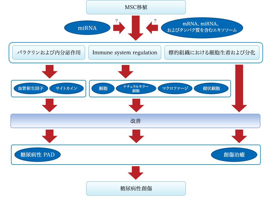
Figure 2: Mechanism of the effect of MSC transplantation on diabetic wounds. Diabetic wound recovery by MSC transplantation from three pathways: first is angiogenesis and secretion of factors and cytokines, second is regulation of the immune system, and third is transplantation and differentiation of cells into tissue constituents. is there. Stem cells can specifically improve the local secretion and expression of angiogenic factors and cytokines that contribute to the improvement of diabetic PAD and diabetes. Stem cells can also regulate the activity of T cells, natural killer cells, macrophages, and dendritic cells, inhibiting infectious and inflammatory responses. In addition, MSCs can differentiate into target tissues and achieve repair. These effects may be associated with miRNAs and MEX. 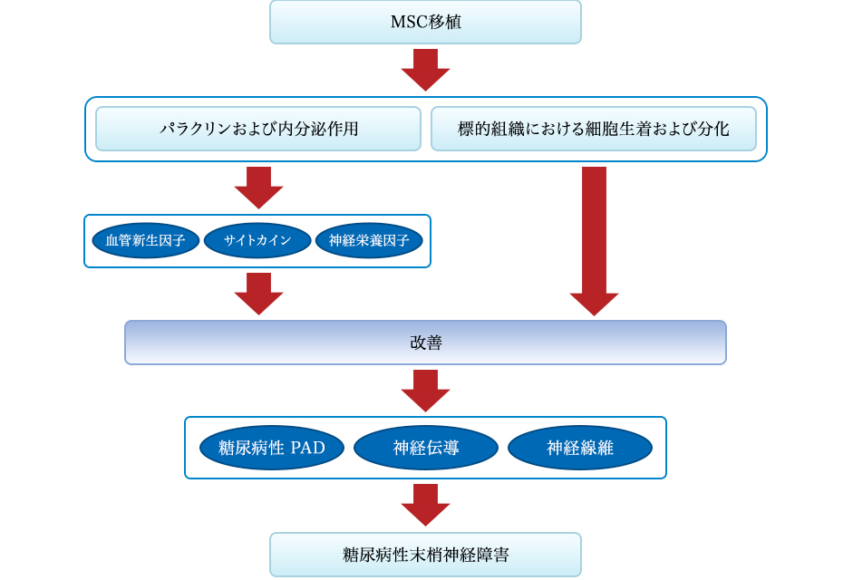
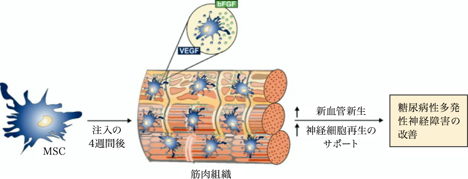
Figure 3: Mechanism of the effect of MSC transplantation on diabetic neuropathy. Mechanisms of recovery effects mediated by stem cell transplantation resulting from two pathways: one is the secretion of angiogenic factors, cytokines and neurotrophic factors, and the other is transplantation and differentiation of cells into tissue constituents.
Stem cells can specifically improve the local secretion and expression of angiogenic factors and cytokines, contribute to the improvement of diabetic PAD and diabetes itself, and lead to the improvement of diabetic neuropathy. Neurotrophins can also improve nerve fiber dysfunction and nerve conduction.
In addition, stem cells can differentiate into target tissues and achieve repair.
kidney failure
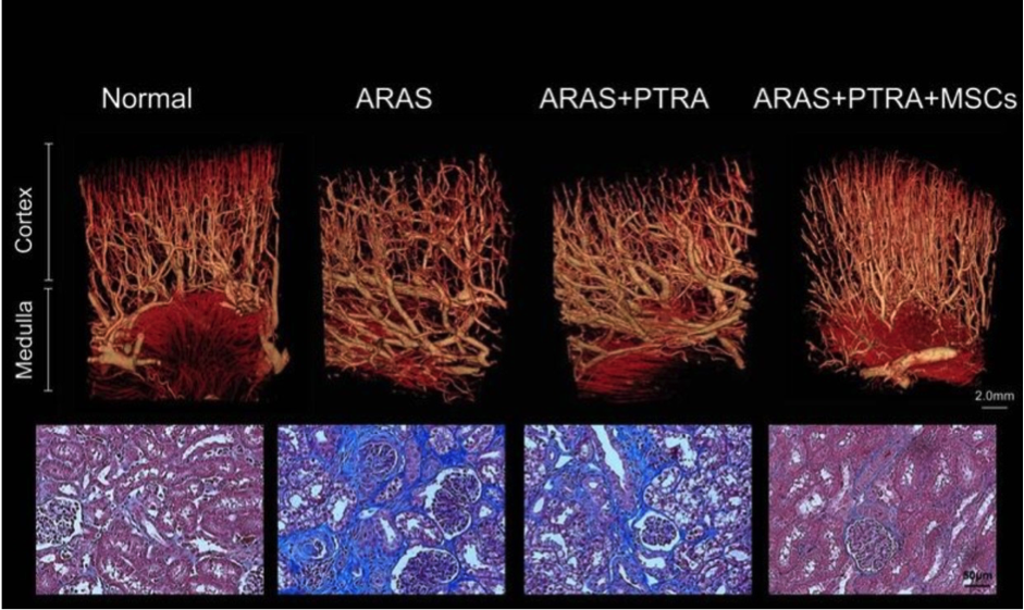
Stenosis-renal microvascular loss and fibrosis were reduced in animals receiving mesenchymal stem cell therapy. Above: A representative microcomputer tomography of the renal segment capturing improved microvascular structure in pigs with atherosclerotic renal artery stenosis undergoing percutaneous transluminal renal angioplasty (PTRA) 3D In the image, adipose tissue-derived mesenchymal stem cells (MSC) were injected intrarenally 4 weeks earlier. Bottom: Representative renal trichrome staining (x40, blue) showing reduced MSC fibrosis in ARAS + PTRA + pigs
Clinical application of MSC: Diabetes
Stem cell transplantation can be a safe and effective treatment for patients with DM. In this series of trials, D34 + HSC therapy achieved the best outcomes for T1DM, while HUCB observed the worst outcomes for T1DM. Diabetic ketoacidosis interferes with the therapeutic effect.
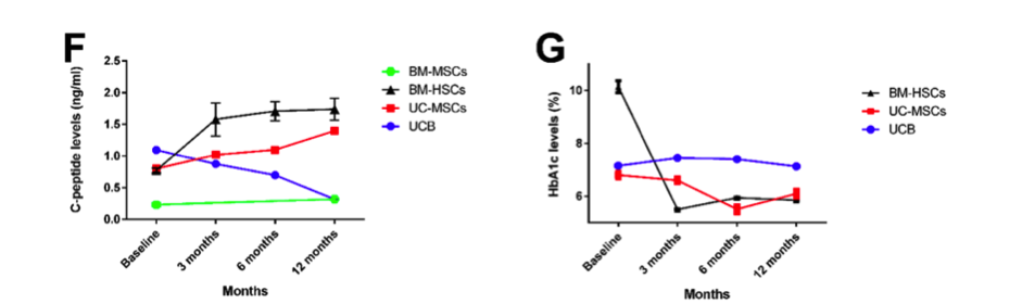
Line graph showing changes in C-peptide and HbA1c levels at baseline, 3, 6, and 12 months after stem cell therapy in T1DM patients. All data are represented as mean ± SEM. **** P <0.0001
The outcome for stem cell therapy for T2DM
Stem cell therapy for type 2 DM.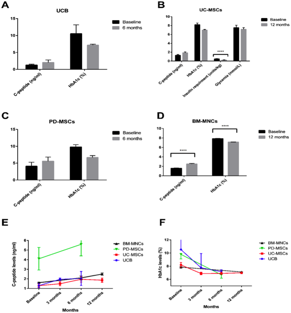
A-D)Bar graph showing baseline after administration of different types of stem cells and changes in C-peptide and HbA1c levels after 12 months. UC-MSC and PD-MSC were injected intravenously (n = 22 and n = 10 respectively), while UCB and BM-MNC were injected intrapancreas (n = 3 and n = 107) (EF) stem cells in T2D. A broken line graph showing changes in C-peptide and HbA1c levels at baseline, 3, 6, and 12 months after treatment.
Citation// PLoS One. 2016 Apr 13;11(4):e0151938. Clinical Efficacy of Stem Cell Therapy for Diabetes Mellitus: A Meta-Analysis. El-Badawy A, El-Badri N.
By disease, hair follicles
Nat Commun. 2012 Apr 17;3:784. doi: 10.1038/ncomms1784.
Fully functional hair follicle regeneration through the rearrangement of stem cells and their niches.
Toyoshima KE1, Asakawa K, Ishibashi N, Toki H, Ogawa M, Hasegawa T, Irié T, Tachikawa T, Sato A, Takeda A, Tsuji T.
Overview
Organ replacement regenerative medicine is said to enable replacement of organs damaged by disease, injury or aging in the foreseeable future. Here we demonstrate fully functional organ regeneration through bioengineering bone and intradermal transplantation of spore germs. The blastoderm and ovule are reconstituted with embryonic skin-derived cells and adult stem cell region-derived cells, respectively. Bioengineered hair follicles develop the correct structure and form proper connections with surrounding host tissues such as epidermis, hindlimb muscles and nerve fibers. Bioengineered hair follicles also exhibit a restored hair cycle and hair head formation through the rearrangement of hair follicle stem cells and their niches. Therefore, this study reveals the potential of adult tissue-derived hair follicle stem cells as a bioengineering organ replacement therapy.
 .
.
(A) Schematic of the method used to produce and transplant bioengineered hair follicle embryos. (B) Phase-difference images of the dorsal skin, tissues, dissociated single cells and bioengineered hair follicle embryos of mouse embryos reconstructed using the organ germination method using nylon threads (arrowheads). Scale, 200 μm.
(c) Histological analysis of isolated tactile hair from adult mice. Macroscopic and H & E stained bristles are shown in the two panels on the left. Dashed lines (red) by macromorphological observation (left) and H & E staining (right) indicate the interface between the bulge and the SB region. The box area on the left panel is H & E stained to show the bulge, and the SB area is shown in a higher magnified view on the right panel. The bulge region includes anti-CD49f (red, left) and anti-CD34 (red, center) antibodies and Hoechst.
It was immunostained with 33258 dye (blue). The black dashed line at high magnification H & E indicates the interface of the epithelium of the hair follicle. IF, funnel; RW, ring; half of the hair follicle. Scale, 100 μm. (d)
Histological and ALP analysis of the valve region of the bristles and the initial culture of DP cells. Hair bulbs (2 on the left) and cultured DP cells (2 on the right) were analyzed by enzymatic staining of ALP. The red dotted line shows the Auber line. Scale ", 100 μm.
(e) Longitudinal section of bioengineered hair during the eruption and anagen process, mediated by an interepithelial tissue connecting plastic device (guided). Corresponding ones are shown as cyst formation with bioengineered follicles 14 days after intradermal transplantation (without guidance). H & E staining of bioengineered hair follicles (top) and fluorescence microscopy (bottom) at 0, 3 and 14 days after transplantation. Scale, 100 μm.
(f) Bioengineered generation, including wound healing on days 0 (left) and 3 (center), hair shaft eruption on days 14 and 37 (right), and growth Macromorphological observation of grown hair during development and growth of the chest (top) and spleen (bottom).
Scale, 1.0 mm.
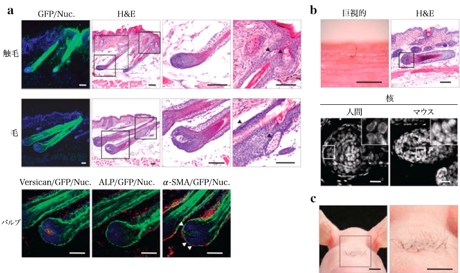
(a) Histological and immunohistochemical analysis of bioengineered hair (upper) and tactile (middle) vesicles. The box area of the low magnification H & E panel is shown at higher magnification in the right panel. Arrows indicate sebaceous glands. Scale, 100 μm. Bioengineered hair follicle hair bulbs are anti
– Immunostained with Versican (lower left) and α-SMA (arrowhead, lower right) antibodies, and also enzymatically stained with ALP. (Bottom center) Scale, 50 μm. (b)
Bioengineered human hair produced by transplantation of biotreated hair follicle embryos was reconstituted with bulge-derived epithelial cells and intact DP of human scalp hair follicles. On the 21st day after transplantation, bioengineered human hair was photographed (microscopic examination) and analyzed by H & E staining. Species identification of bioengineered hair follicles was analyzed according to nuclear morphological features (right panel). The enclosed area of the inset is shown at high magnification. Scale, microscope 500 μm, H & E 100 μm, nuclear staining 20 μm. (c)
High-density intradermal transplantation of bioengineered folliculous pathogens. A total of 28 independent bioengineered hair follicle germs were transplanted into the cervical skin of mice and showed dense hair growth 21 days after transplantation. Scale, 5 mm.
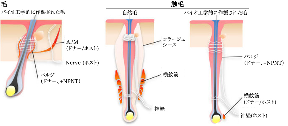
Bioengineered hairs and tactile hairs were linked to other tissues such as nerve fibers, traction and streak muscles derived from host or donor cells. Bioengineered hairs bound to smooth muscle as a result of regeneration of the bulge region expressing NPNT as well as natural masses. Neither NPNT expression nor smooth muscle binding was detected in the bioengineered hair bulge region.
Quote/ Fully functional hair follicle regeneration through the rearrangement of stem cells and their niches. Koh-ei Toyoshima, Kyosuke Asakawa, Naoko Ishibashi, Hiroshi Toki, Miho Ogawa, Tomoko Hasegawa, Tarou Irié, Tetsuhiko Tachikawa, Akio Sato, Akira Takeda & Takashi Tsuji. Nature Communications 3, Article number: 784 (2012)
doi:10.1038/ncomms1784
Other diseases, Parkinson
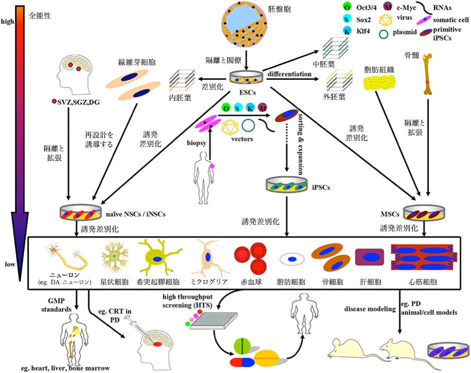
Schematic of the induction, differentiation and application of stem cells currently available in PD studies and treatments. The above stem cells can be divided into four categories: ESC, NSC, MSC, and iPSC, with gradually diminishing totipotency.
(1) ESC, which is mainly derived from the inner cell mass of blastocysts, can simultaneously differentiate into endoderm, mesoderm and ectoderm under normal circumstances. In some cases, ESCs can also be induced to differentiate into NSCs and MSCs.
(2) NSCs isolated directly from specific brain niches or reprogrammed from fibroblasts can differentiate the nervous system into neurons and almost all glial cells. (3)
MSCs are primarily derived from mesenchymal tissue and can differentiate into almost any cell of mesoderm origin. Notably, MSCs can be induced to differentiate into DA neurons even under certain combinations of induction protocols. (Four)OSKM(Oct3 / 4、Sox2、Klf4、およびc-Myc)を導入することにより、成人のヒト体細胞(線維芽細胞など)から再分化させることができるiPSCsは、多系譜分化能を有する有望な幹細胞源である。GMP標準に基づいて、上記の幹細胞および最終分化細胞を、疾患モデル、薬物スクリーニング、およびCRTの実施に適用するために、さらに選別し、精製し、そして拡大することができる。例えば、ESC、MSC、NSC、およびDAニューロンは、以下で使用される。(i)PDモデルの準備(ii)潜在的な薬物スクリーニング;(iii)PDのCRT治療Front.
Aging Neurosci., 31 May 2016. A Compendium of Preparation and Application
of Stem Cells in Parkinson’s Disease: Current Status and Future Prospects.
Yan Shen, Jinsha Huang